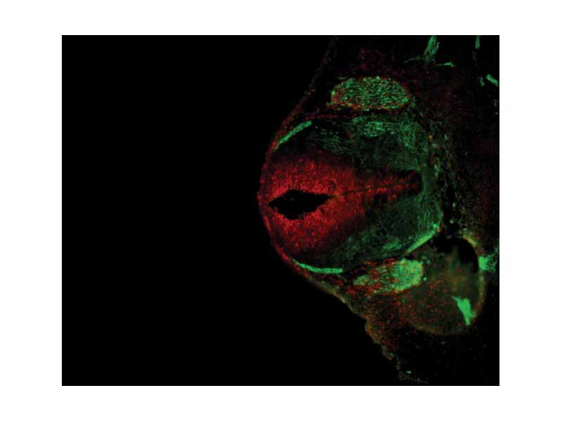
Antibody stain against Neurofilament (green) and Ki 67 (red) in a Mouse embryo 12.5 days after fertilization. The cells expressing neurofilaments are in the dorsal root ganglions shown in green while proliferating cells are in the ventricular zone in the neural tube and colored red.
Click this LINK to visit the original image and attribution information. Right click on the image to save the 800px teaching JPEG.